

Chest Pain: Is It Ever Normal?
Pain in your chest can be frightening because your first thought is that your heart is in danger. It may or may not be. Keep reading to learn what causes chest pain and when to seek medical care.
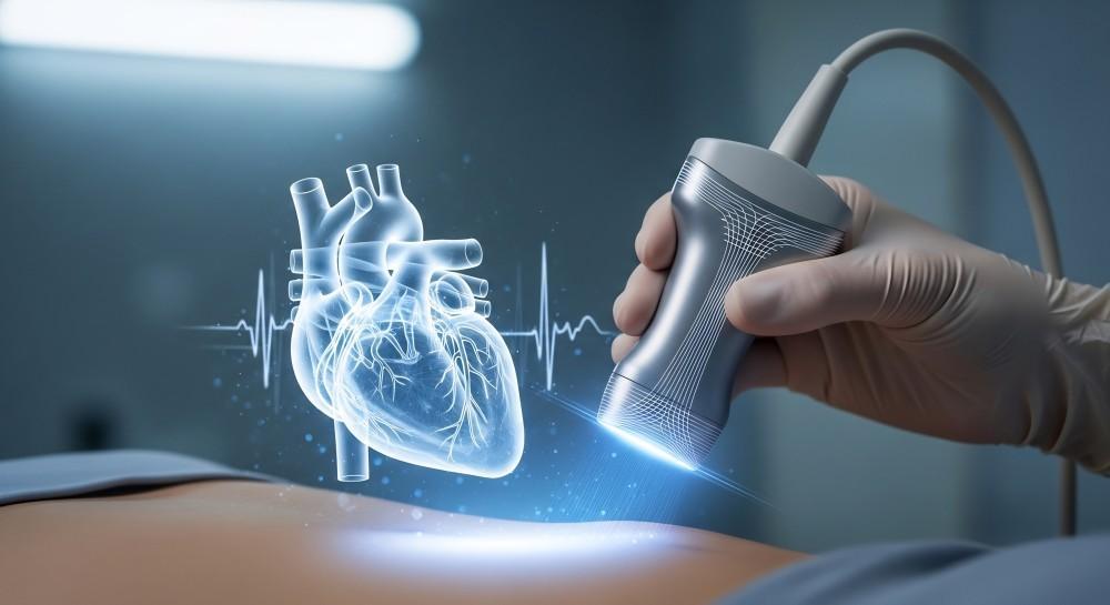
When patients visit us for chest discomfort, shortness of breath, or an irregular heartbeat, a cardiac diagnostic evaluation is often the initial step in diagnosing the cause.
Two of the most common non-invasive cardiac diagnostic tests are an electrocardiogram (EKG) and an echocardiogram. While both tests evaluate cardiovascular function, they examine entirely different aspects of the heart.
At South Palm Cardiovascular Associates in Boynton Beach, FL, our board-certified cardiologists and specialists use both modalities to accurately diagnose conditions.
An EKG records electrical signals, while an echo uses sound waves to create moving pictures of cardiac structures.
Understanding how each test works can relieve anxiety before an appointment. Here’s what our cardiologists want you to know.
An EKG records the electrical currents that trigger each heartbeat. During this painless test, a technician attaches small adhesive patches with sensors to your chest, arms, and legs. These sensors measure the timing and strength of electrical impulses as they travel through cardiac tissue.
The procedure takes between five and ten minutes. You lie flat and remain still while the machine records the electrical tracings on a graph.
An EKG detects:
Because the test takes only minutes, our team uses it as an efficient first-line assessment when patients experience palpitations, dizziness, or chest tightness.
An echocardiogram is a specialized ultrasound examination that captures moving images of your heart chambers, valves, and surrounding blood vessels. While an EKG tracks electrical signals, an echo assesses physical architecture and mechanical pumping function.
During a standard transthoracic echocardiogram, you lie on an examination table while a sonographer moves a handheld transducer across your chest. High-frequency sound waves bounce off cardiac tissues, transmitting signals to a computer that creates real-time video clips of the beating heart. The appointment generally takes between thirty and forty-five minutes.
An echocardiogram evaluates:
An echocardiography is the primary imaging modality for evaluating heart failure and valvular disease. It provides detailed measurements of ejection fraction and blood flow velocities.
Although patients often confuse these tests due to similar names, several differences set them apart:
An EKG evaluates electrical conduction, whereas an echocardiogram assesses muscular structures, valves, and blood flow.
An EKG takes under ten minutes, while an echocardiogram takes roughly thirty to forty-five minutes.
An EKG shows if rhythm problems exist; an echo reveals whether mechanical damage or muscular weakness caused those rhythm problems.
Neither test requires invasive needles or sedation, though an echo requires acoustic gel applied directly to the chest wall.
Our experts frequently order both evaluations together. An irregular rhythm on an EKG might prompt an echocardiogram to determine whether valve dysfunction or chamber enlargement is causing the abnormal beat.
If you experience irregular heartbeats, unexplained fatigue, or chest pain, getting an accurate diagnosis is the first step towards alleviating these symptoms. Contact the expert cardiology team at South Palm Cardiovascular Associates to schedule your appointment today.